A successful impression does not depend only on the material or the scanner: it depends first of all on what is visible at the moment of recording. If the margin is drowned in gingival fluid or hidden by the marginal gingiva, the restoration that results will at best be an interpretation. Sulcus management is a prosthetic step in its own right.
An invisible margin is an interpreted margin
At the lab, tracing the margin is the first operation performed on the model or the file. When it is readable on only part of the circumference, the dental technician has two options: interpolate the missing portion, or call you back. In the first case, the seal will be approximate exactly where clinical control is most difficult; in the second, the appointment is postponed.
The goal is simple to state: a sharp, exposed margin over 360°, with enough lateral space for the impression material or the scanner beam to reach the bottom of the sulcus.
Gingival retraction techniques
The single cord
A single cord, impregnated or not, is suitable for juxtagingival margins and healthy, shallow sulci. It is placed without force, left in place according to the manufacturer's instructions, then removed just before injection. Its main drawback is that it lifts out when the sulcus is wide.
The double-cord technique
It remains the reference for intrasulcular margins. A first, thin cord is left in place during the impression: it ensures hemostasis and maintains vertical opening. A second, thicker cord is placed on top and removed at the time of injection, freeing up lateral space. It is the most reproducible technique on a thin periodontium.
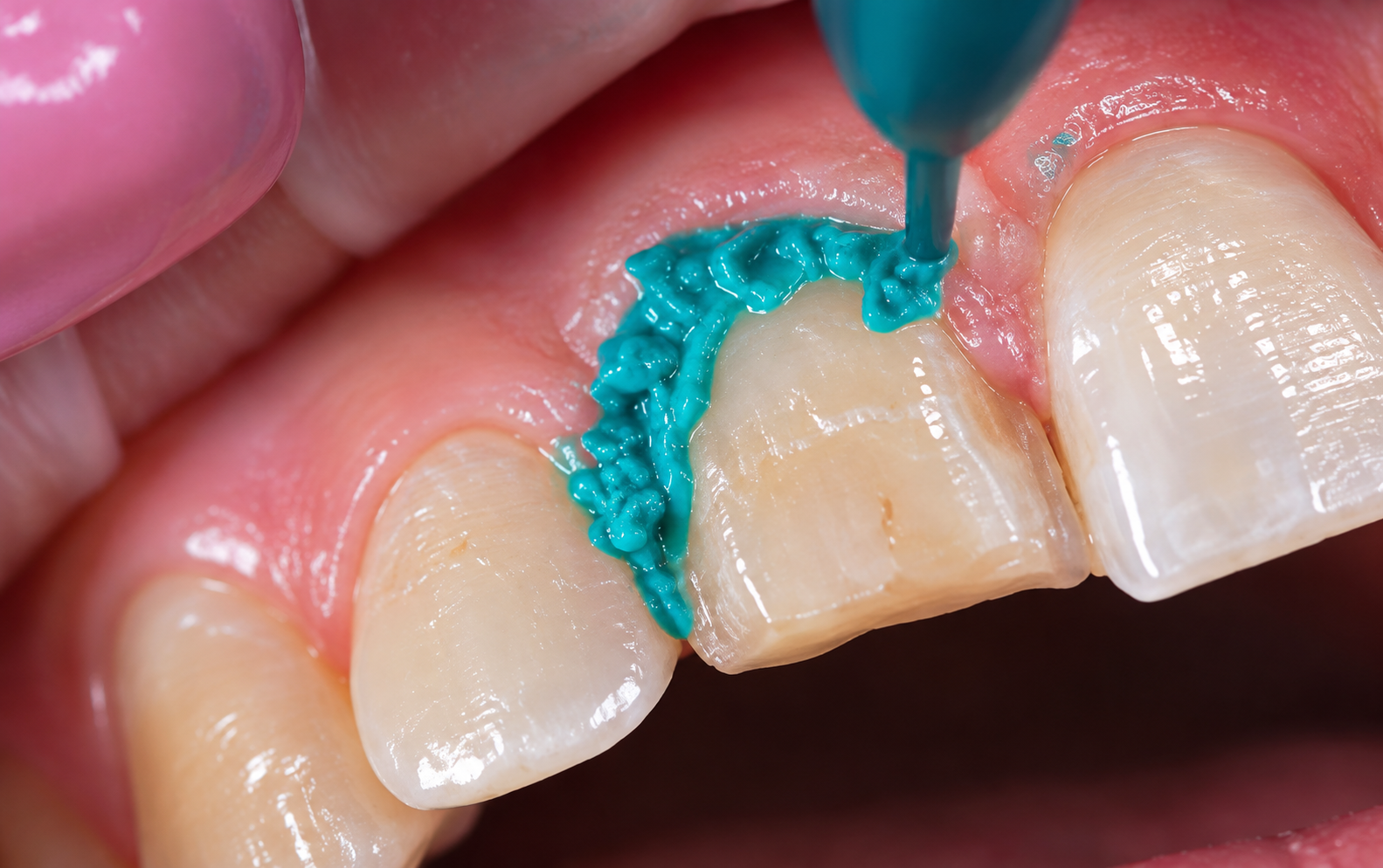
Retraction pastes
Injected and then rinsed, they are less traumatic and faster, and work well for juxtagingival margins. Their lateral opening capacity is, however, lower than that of a cord, which limits their usefulness on deep margins.
Surgical approaches
When the sulcus is inflamed, the margin encroaches on the biological width or the gingiva is hypertrophic, no retraction technique will compensate. A minimal gingivectomy, a crown lengthening or simply a healing period after periodontal treatment will restore workable conditions. It is better to postpone the impression than to record it in poor conditions.
Which technique for which situation
| Clinical situation | Preferred technique | Points of attention |
|---|---|---|
| Supragingival margin, healthy periodontium | No retraction, or retraction paste | Check for the absence of fluid before injecting |
| Juxtagingival margin | Single cord or retraction paste | Respect the manufacturer's placement time |
| Intrasulcular margin, thin periodontium | Double cord | Place without excessive pressure to avoid recession |
| Bleeding or inflamed sulcus | Periodontal treatment, then postpone the impression | Treat the cause before recording |
| Hypertrophic gingiva, hidden margin | Minimal surgical correction | Wait for the gingival scallop to stabilize |
A reproducible protocol
- Check the periodontal condition and postpone if the gingiva bleeds on probing.
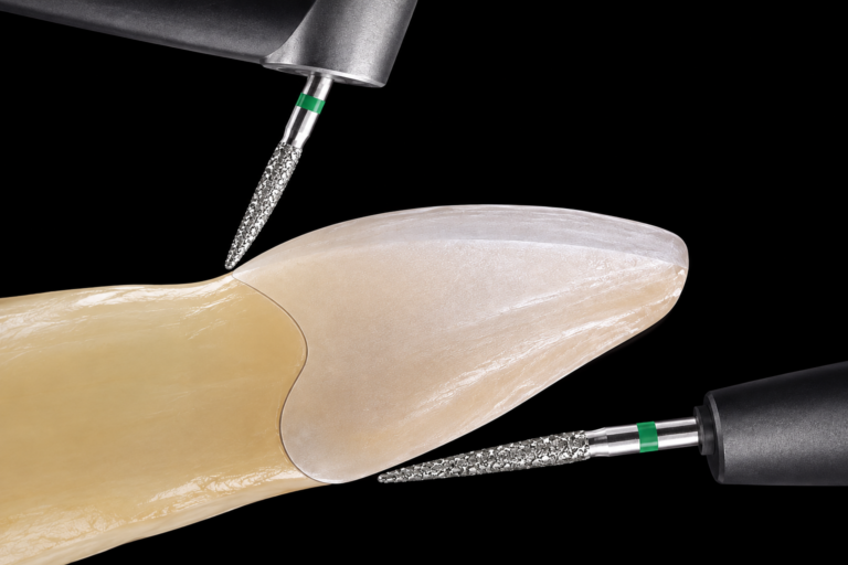
- Finish and polish the margin before any retraction: an irregular margin will remain unreadable, even when perfectly exposed.
- Choose the technique according to the depth of the margin and the gingival biotype.
- Place the cord in small sections, pressing against the tooth rather than the gingiva.
- Respect the placement time indicated by the manufacturer, then moisten before removal so as not to restart bleeding.
- Dry without desiccating, then inject immediately and continuously at the bottom of the sulcus.
- Check the impression or the file on screen, margin by margin, before letting the patient go.
What the lab can, and cannot, compensate for
A slight lack of material over a short area can be smoothed out with no clinical consequence. However, a margin missing over several millimeters, a fold of retracted material or a bubble located exactly on the chamfer cannot be corrected: they can only be guessed. The restoration may be esthetic, but the seal will not be the one you prepared.
How Dune Dental supports you
On receipt, every impression — physical or digital — undergoes a margin readability check before going into production. When an area is uncertain, we send you an annotated screenshot of the area concerned: you then decide whether to approve the tracing or redo the recording, with full knowledge of the facts, rather than discovering it at the try-in. [To be completed: Dune Dental's response time for an impression check] [To be completed: channel used to send the annotated screenshots]
FAQ
Impregnated or dry cord?
Both work. Cords impregnated with an astringent improve hemostasis, which is useful on a reactive sulcus; on a healthy periodontium, a well-placed dry cord is often enough. In every case, the placement time follows the manufacturer's instructions.
Does the laser change the approach?
It allows exposure and coagulation in a single step, which is convenient on a bleeding sulcus, but requires mastery of the settings: excessive irradiation can cause recession. The prosthetic logic, however, remains the same.
Do digital impressions make retraction unnecessary?
No, quite the opposite. The scanner only records what it sees: it does not move the gingiva and cannot see through blood or saliva. Access requirements are at least as strict as for a physical impression.
How long should you wait after a gingivectomy?
The timing depends on the extent of the procedure and the biotype, and is generally counted in weeks. The benchmark remains the stabilization of the gingival scallop: recording too early risks seeing the margin shift after seating.