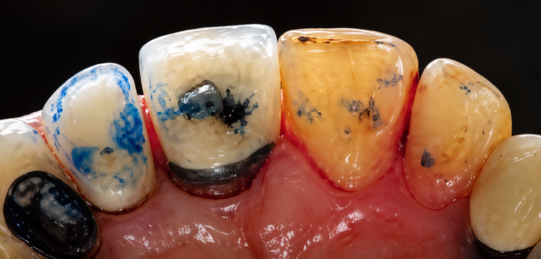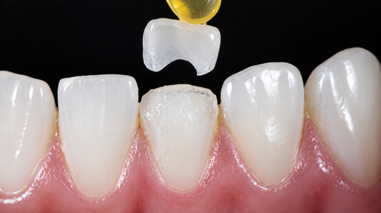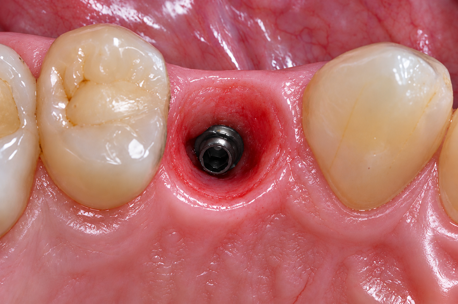
Red gingiva that bleeds on probing and does not settle despite good plaque control, around a crown placed a few months earlier: the situation is common, and often blamed on the patient's hygiene. In a significant share of cases, the cause is prosthetic — and the emergence profile is the prime suspect.
What the emergence profile covers
The emergence profile is the portion of the restoration between the prosthetic margin and the contact point, where the restoration passes through the sulcus and then emerges into the oral cavity. A narrow area, invisible once the restoration is seated, it nevertheless determines both the patient's access for cleaning and the stability of the surrounding tissue.
A favorable profile is most often slightly concave, then straight as it leaves the sulcus: it accompanies the tissue without pushing it away. As soon as the restoration becomes convex in this area, it exerts permanent pressure on the marginal gingiva, which responds with chronic inflammation or recession.
The prosthetic causes of inflammation
Overcontouring
This is the most frequent cause. An overly convex profile creates a retention area where the patient can no longer pass floss or an interdental brush: inflammation sets in and persists regardless of their motivation. A few tenths of a millimeter are enough to make the difference.
Surface finish and the margin seal
Insufficiently glazed ceramic, adjusted chairside without repolishing, or a margin with a step provide a surface that favors biofilm accumulation. This is all the more critical intrasulcularly, where cleaning is out of the patient's reach.
Excess cement
A residue left in the sulcus sustains an inflammation that mimics localized periodontitis. The risk increases with the depth of the margin: it is a further argument in favor of juxtagingival or very slightly intrasulcular margins, and of a radiographic check after cementation when in doubt.
Encroachment on the biological width
When the margin is placed too apically, the tissue response is predictable: persistent inflammation, then recession until sufficient space is restored. No change to the profile will correct this situation; it is the position of the margin that must be revised, most often surgically.
From clinical sign to probable cause
| Observed sign | Prosthetic cause to suspect | Possible correction |
|---|---|---|
| Bleeding localized to a single surface | Overcontouring or excess cement on that surface | Radiographic check, profile adjustment or removal |
| Floss that shreds or does not pass | Inadequate contact point or step at the margin | Rework of the contact point and repolishing |
| Inflammation around several crowns | Systematically convex profiles, insufficient access for cleaning | Review the design with the lab |
| Progressive recession, margin now visible | Encroachment on the biological width | Surgical reassessment, then a new prosthesis |
| Bluish gingiva, dark line | Leakage at the margin or framework showing through | Revision of the margin, change of material |
A simple diagnostic approach
- Objectify: probing, bleeding, photograph of the area and periapical radiograph.
- Check whether floss and an interdental brush pass: being unable to clean points immediately to overcontouring or an inadequate contact point.
- Look for excess cement, particularly if the inflammation appeared in the weeks following seating.
- Assess the distance between the margin and the bone crest before considering an adjustment.
- Decide: adjustment and repolishing, removal and remake, or prior periodontal treatment.
Prevention starts at the design stage
Prevention happens upstream, when the restoration is designed. Three elements contribute: margins placed at the shallowest level compatible with esthetics and retention, a profile designed from a recording that truly captures the sulcus, and checking that cleaning instruments pass at the try-in, before final luting. Provisionalization is also an excellent test: calm tissue around the provisional announces calm tissue around the definitive restoration.
How Dune Dental supports you
The emergence profile is one of the points we systematically check at the design stage, before milling. When the sulcus recording is insufficient to design a reliable profile, we flag it rather than guess. We can also start from a provisional approved in the mouth in order to reproduce a profile that has already proven itself clinically — the safest method in esthetic areas and around implants. [To be completed: how to send a provisional or a customization impression to Dune Dental] [To be completed: technical team contact for an opinion on an ongoing case]
FAQ
Can an emergence profile be adjusted chairside?
Yes, provided you remain conservative and repolish carefully: ceramic that is adjusted and not repolished worsens the problem you are trying to solve. Beyond a few tenths of a millimeter, remaking the restoration is more reliable.
Does the material influence the gingival response?
The quality of the surface finish and the margin seal matters more than the nature of the material. That said, properly glazed and polished ceramic is well tolerated by the tissues, which makes it a comfortable choice in contact with the gingiva.
How do you tell a prosthetic cause from periodontitis?
Location is the best clue: inflammation limited to the area around a single restoration, on an otherwise healthy periodontium, points to a prosthetic cause. A full periodontal assessment remains necessary in case of generalized involvement.
Should a crown be removed in case of excess cement?
Not systematically. If the residue is accessible, removing it and cleaning the area are often enough. If it is deep and the inflammation persists, removal becomes the most predictable solution.